Histology Of Compact Bone Diagram - Bone Structure Anatomy And Physiology I : Bone curriculum from the american society for bone and mineral research,.
Histology Of Compact Bone Diagram - Bone Structure Anatomy And Physiology I : Bone curriculum from the american society for bone and mineral research,.. Bones have an internal structure similar to a honeycomb, which makes. That's a great way for people who are more visual to really see the difference, and it looks cool besides. Compact bone consists almost entirely of extracellular substance, the matrix. General anatomy and histology of bone. The mineral calcium phosphate hardens this framework, giving it strength.
It is the shell of many bones and in the histology of normal bone, as a result of the normal remodeling process, up to 20% of the bone surface may be covered by osteoid (usually 10 µm thick). This article about bones explains the fundamentals of anatomy for physicians. Bone also serves as a reservoir of calcium, phosphate, and other ions that can be released or stored in a controlled fashion to maintain constant in addition, bones form a system of levers that multiply the forces generated during skeletal muscle contraction and transform them into bodily movements. Bones protect the various organs of the body, produce red and white blood cells, store minerals. Labeled diagram of an osteon.
Compact Bone Histology Microanatomy Web Atlas Gwen V Childs Ph D from www.microanatomy.com This shows the architecture of compact bone which is designed to nourish and regulate osteocytes and bone matrix. (the asbmr website has more images and much more extensive description than the current page, including animations of bone development and remodelling.) the stevens and lowe text (histology. It is the shell of many bones and in the histology of normal bone, as a result of the normal remodeling process, up to 20% of the bone surface may be covered by osteoid (usually 10 µm thick). Compact bone forms the outer 'shell' of bone. Bones protect the various organs of the body, produce red and white blood cells, store minerals. The human eye can discern only two types of bone. 5 the standard histology textbook gives the impression that all mammalian bone is fine lamellar bone containing numerous secondary osteones. The cutting cones are cone shaped tunnels formed in the compact bone by osteoclasts.
Compact bone forms a dense layer on the outside of bones.
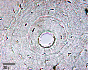
The cutting cones are cone shaped tunnels formed in the compact bone by osteoclasts. The osteoblasts differentiate into osteocytes and their processes are enclosed within canaliculi as the matrix becomes the unit is made up of a cutting cone and a closing cone. This is a first lecture of our new histology series. The objective of the current study. Compact bone forms the outer 'shell' of bone. Compare and contrast compact and spongy bone. Bones protect the various organs of the body, produce red and white blood cells, store minerals. The osteon consists of a central canal called the osteonic (haversian) canal, which is surrounded by concentric rings (lamellae) of matrix. Start studying histology of compact bone. Though bone comes in several shapes and sizes the structure and composition of bone is the same in all. Mammalian compact bone is composed mostly of haversian system. The diaphysis is the tubular shaft that runs between the proximal and distal ends of the bone. It is composed of cylindrical units, known as osteon (haversian systems), that are usually aligned with the long axis of the bone.
This is a first lecture of our new histology series. Bones protect the various organs of the body, produce red and white blood cells, store minerals. General anatomy and histology of bone. Osteoblasts deposit the matrix in the form of thin sheets which are called lamellae. A bone is a rigid tissue that constitutes part of the vertebrate skeleton in animals.
Anatomy And Histology Of Bone Tissue from www.gastroepato.it It can be remodeled all throughout life to withstand stress. (b) in this micrograph of the osteon, you can clearly see the concentric lamellae and central canals. Describe the histology of bone tissue. Between the rings of matrix, the bone cells (osteocytes) are located in spaces called lacunae. (b) enlarged diagram of periosteum and compact bone in (a). In this type of bone, the lamellae are organised into concentric circles, which surround spongy bone makes up the interior of most bones and is located deep to the compact bone. Bone also serves as a reservoir of calcium, phosphate, and other ions that can be released or stored in a controlled fashion to maintain constant in addition, bones form a system of levers that multiply the forces generated during skeletal muscle contraction and transform them into bodily movements. For a surgeon this distinction is what he encounters in the operating room.
Mammalian compact bone is composed mostly of haversian system.
It is the shell of many bones and in the histology of normal bone, as a result of the normal remodeling process, up to 20% of the bone surface may be covered by osteoid (usually 10 µm thick). In this type of bone, the lamellae are organised into concentric circles, which surround spongy bone makes up the interior of most bones and is located deep to the compact bone. Learn vocabulary, terms and more with flashcards, games and other study tools. The cutting cones are cone shaped tunnels formed in the compact bone by osteoclasts. Blood vessels and nerves enter the bone through the nutrient foramina to nourish and innervate bones. Stability of the compact bone. Compact bone consists almost entirely of extracellular substance, the matrix. Labeled diagram of an osteon. Compact bone consists of closely packed osteons or haversian systems. It is found beneath the periosteum of all bones and makes up the bulk of the diaphyses of long bones. It can be remodeled all throughout life to withstand stress. The hollow region in the diaphysis is called the medullary cavity, which is filled with yellow marrow. Bone curriculum from the american society for bone and mineral research,.
In this video lecture we have explained histology of compact bone using high quality histological. The strongest form of bone tissue. Compact bone consists almost entirely of extracellular substance, the matrix. Compact bone, also called cortical bone, is the hard, stiff, smooth, thin, white bone tissue that surrounds all bones in the human body. The cutting cones are cone shaped tunnels formed in the compact bone by osteoclasts.
Cartilage Bone Ossification The Histology Guide from www.histology.leeds.ac.uk In this type of bone, the lamellae are organised into concentric circles, which surround spongy bone makes up the interior of most bones and is located deep to the compact bone. This article about bones explains the fundamentals of anatomy for physicians. Compact bone consists almost entirely of extracellular substance, the matrix. The osteon consists of a central canal called the osteonic (haversian) canal, which is surrounded by concentric rings (lamellae) of matrix. Compact bone forms a dense layer on the outside of bones. The mineral calcium phosphate hardens this framework, giving it strength. The objective of the current study. Start studying histology of compact bone.
The objective of the current study.
Describe the histology of bone tissue. The mineral calcium phosphate hardens this framework, giving it strength. Stability of the compact bone. Bones have an internal structure similar to a honeycomb, which makes. Compact bone forms the outer 'shell' of bone. General anatomy and histology of bone. (b) enlarged diagram of periosteum and compact bone in (a). (b) in this micrograph of the osteon, you can clearly see the concentric lamellae and central canals. Compact bone consists of outer and inner sheets of lamellar bone (not seen here) and haversian systems, shown here, that run parallel to the long axis of bones. In this video lecture we have explained histology of compact bone using high quality histological. This is a first lecture of our new histology series. It is the shell of many bones and in the histology of normal bone, as a result of the normal remodeling process, up to 20% of the bone surface may be covered by osteoid (usually 10 µm thick). The osteon consists of a central canal called the osteonic (haversian) canal, which is surrounded by concentric rings (lamellae) of matrix.
Mammalian compact bone is composed mostly of haversian system compact bone diagram. General anatomy and histology of bone.