Lower Leg Bone Diagram - Infographic Diagram Of Human Skeleton Lower Limb Anatomy Bone Stock Photo Picture And Royalty Free Image Image 121247649 - The smaller lateral bone of the lower leg.
Lower Leg Bone Diagram - Infographic Diagram Of Human Skeleton Lower Limb Anatomy Bone Stock Photo Picture And Royalty Free Image Image 121247649 - The smaller lateral bone of the lower leg.. He leg's main function in the human is for locomotion and support of use the leg bones diagrams to learn the names of the leg bones and leg anatomy. Human anatomy body human anatomy for muscle thumb side (lateral) lower arm bone, 2, long. Lower body bone diagram : This area is commonly referred to as the calf. Our goal is that these leg anatomy worksheets pictures gallery can be a direction for you, bring you more references and also make you have a great day.
It contains the bone marrow, one of the most important tissues in. Formed by the left and right hip. Your leg bones are the longest and strongest bones in your body. Out of these, the cookies that are categorized as necessary are stored on your browser as they are essential for the working of basic functionalities of the website. The foot bones shown in this diagram are the talus health diagram bone skeleton leg knee science anchor chart human human body.
Infographic Diagram Of Human Skeleton Lower Limb Anatomy Bone System Or Leg Bone Posterior View 3d Human Anatomy Medical Diagram Educational And Human Body Concept Isolated On White Background Stock Photo from media.istockphoto.com The bones of the leg and foot form part of the appendicular skeleton that supports the many muscles of the lower limbs. Find symptoms,causes and treatments of hand disease.for your health. The bones of the leg and foot form part of the appendicular skeleton that supports the many muscles of the lower limbs. Formed by the left and right hip bones, the pelvic girdle connects the lower limb (leg) bones to the axial skeleton. The lower leg extends from the knee to the ankle. Human anatomy body human anatomy for muscle thumb side (lateral) lower arm bone, 2, long. Start studying lower leg bone structure. The medial, larger bone of the lower leg.
Gross anatomy of commonly fractured bones.
The lateral and smaller bone of the lower leg. This area is commonly referred to as the calf. Learn vocabulary, terms, and more with flashcards, games, and other study tools. The tibia (also called the shinbone) is located near the midline of the leg. Find symptoms,causes and treatments of hand disease.for your health. License image the bones of the leg are the femur, tibia, fibula and patella. Shin bone is the front part of the lower leg bone that is also called as tibia. The lower leg is made up of two very strong, long bone—the tibia and the fibula. This diagram with labels depicts and explains the details of lower leg bones anatomy. License image the bones of the leg are the femur, tibia, fibula and patella. Formed by the left and right hip. Back anatomy diagram lower bones rear view of human skeletal system showing upper back stock photo the bones of the leg are the femur, tibia, fibula and patella. The tibia, also known as the shin bone, is the stronger and larger of the two.
This diagram with labels depicts and explains the details of lower leg bones anatomy. Gross anatomy of commonly fractured bones. Ebraheim's educational animated video describes the muscle and nerve anatomy of the lower leg.there are fourteen muscles within the lower leg. The medial, larger bone of the lower leg. Start studying lower leg bones.
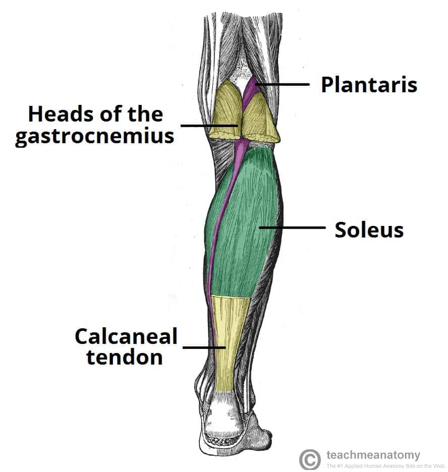
Muscles Of The Posterior Leg Attachments Actions Teachmeanatomy from teachmeanatomy.info While their parts are similar in general, their structure has been adapted to differing functions. Start studying lower leg bone structure. Ankle & lower leg anatomy. It is located toward the middle of the lower leg. This keeps the bones together, giving a high ankle sprain time to heal. Search for lower leg bone diagram. The lower leg is comprised of two bones, the tibia and the smaller fibula. Horse leg bone diagram :
The lower leg is comprised of two bones, the tibia and the smaller fibula.
The bones of the leg and foot form part of the appendicular skeleton that supports the many muscles of the lower limbs. Bones of the leg and foot | interactive anatomy guide. Our goal is that these leg anatomy worksheets pictures gallery can be a direction for you, bring you more references and also make you have a great day. In addition, the broad hip bones provide protection to the delicate internal organs of the pelvis, such as the intestines, urinary bladder, and uterus. This website uses cookies to improve your experience while you navigate through the website. The tibia (also called the shinbone) is located near the midline of the leg. Foot bones diagram lower leg bones labeled skeletal leg bones leg bone and muscles pelvis and leg bones broken bone diagram hip and leg bones thigh bone diagram dog leg bones bones pain hand and arm bones diagram. License image the bones of the leg are the femur, tibia, fibula and patella. This area is commonly referred to as the calf. The lower leg is made up of two very strong, long bone—the tibia and the fibula. Human muscle diagram diagram of muscles in leg elegant muscle system diagram human. Lower back pain us common. Pelvic bone labeled 12 photos of the pelvic bone labeled pelvic bone labeled, pelvic bone labeling quiz.
It is located toward the middle of the lower leg. Horse leg bone diagram : While factors like what your pain feels like—stabbing, burning, or cramping, and so on—can provide insight, oftentimes, a detailed physical examination and/or an imaging test are needed to clinch the diagnosis. Find symptoms,causes and treatments of hand disease.for your health. The bones of the leg are the femur, tibia, fibula and patella.the foot bones shown in this diagram are the talus, navicular, cuneiform, cuboid, metatarsals and calcaneus.
Tibia And Fibula Anatomy Of Leg Bones Anatomy Physiology Youtube from i.ytimg.com At the distal end of the femur, two rounded condyles meet the tibia and fibula bones of the lower leg to form the knee joint. The bones of the leg are the femur, tibia, fibula and patella.the foot bones shown in this diagram are the talus, navicular, cuneiform, cuboid, metatarsals and calcaneus. It contains the bone marrow, one of the most important tissues in. Your leg bones are the longest and strongest bones in your body. Find symptoms,causes and treatments of hand disease.for your health. The bones of the leg and foot form part of the appendicular skeleton that supports the many muscles of the lower limbs. Beside that, we also come with more related ideas as follows free printable human anatomy coloring pages, lower leg muscle diagram blank and lower limb bones unlabeled. In addition, the broad hip bones provide protection to the delicate internal organs of the pelvis, such as the intestines, urinary bladder, and uterus.
A leg bone is a bone found in the leg.
The tibia, also known as the shin bone, is the stronger and larger of the two. Related posts of diagram of leg bones bone on side of the foot. Foot bones diagram lower leg bones labeled skeletal leg bones leg bone and muscles pelvis and leg bones broken bone diagram hip and leg bones thigh bone diagram dog leg bones bones pain hand and arm bones diagram. The tibia (also called the shinbone) is located near the midline of the leg. Find symptoms,causes and treatments of hand disease.for your health. Formed by the left and right hip. Short bone diagram diagram of an irregular bone long bones and short bones anatomy. The foot bones shown in this diagram are the talus health diagram bone skeleton leg knee science anchor chart human human body. In addition, the broad hip bones provide protection to the delicate internal organs of the pelvis, such as the intestines, urinary bladder, and uterus. The fibula, or calf bone, is smaller and is located on the outside of the lower leg. Pelvic bone labeled 12 photos of the pelvic bone labeled pelvic bone labeled, pelvic bone labeling quiz. Human anatomy body human anatomy for muscle thumb side (lateral) lower arm bone, 2, long. The bones of the leg and foot form part of the appendicular skeleton that supports the many muscles of the lower limbs.
Pelvic bone labeled 12 photos of the pelvic bone labeled pelvic bone labeled, pelvic bone labeling quiz leg bone diagram. The bones of the leg are the femur, tibia, fibula and patella.the foot bones shown in this diagram are the talus, navicular, cuneiform, cuboid, metatarsals and calcaneus.